


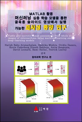

머신러닝 심층 학습 모델을 통한 골육종 슬라이드 영상에서 실행 가능한 괴사성 종양연구
- 저자Harish Babu Arunachalam, Rashika Mishra,Ovidiu Daescu, Kevin Cederberg,Dinesh Rakheja,외
- 출판사아진
- 출판일2020-07-12
- 등록일2020-12-21
- SNS공유


- 파일포맷PDF
- 파일크기20MB
- 공급사YES24
-
지원기기
PC
PHONE
TABLET
프로그램 수동설치
전자책 프로그램 수동설치 안내
아이폰, 아이패드, 안드로이드폰, 태블릿,
보유 1, 대출 0,
예약 0, 누적대출 4, 누적예약 0
책소개
Pathological estimation of tumor necrosis after chemotherapy is essential forpatients with osteosarcoma. This study reports the first fully automated tool to
assess viable and necrotic tumor in osteosarcoma, employing advances in
histopathology digitization and automated learning. We selected 40 digitized whole
slide images representing the heterogeneity of osteosarcoma and chemotherapy
response. With the goal of labeling the diverse regions of the digitized tissue into
viable tumor, necrotic tumor, and non-tumor, we trained 13 machinelearning
models and selected the top performing one (a Support Vector Machine) based on
reported accuracy. We also developed a deep-learning architecture and trained it
on the same data set. We computed the receiver-operator characteristic for
discrimination of nontumor from tumor followed by conditional discrimination of
necrotic from viable tumor and found our models performing exceptionally well. We
then used the trained models to identify regions of interest on image-tiles
generated from test whole slide images. The classification output is visualized as a
tumor-prediction map, displaying the extent of viable and necrotic tumor in the
slide image. Thus, we lay the foundation for a complete tumor assessment pipeline
from original histology images to tumor-prediction map generation. The proposed
pipeline can also be adopted for other types of tumor.
목차
제 1편 : MATLAB 기본편1. MATLAB 기본사용편 ···················· 003
1.1 MATLAB 시작하기 ·························· 003
명령창(command Window)에서의 입력 005
도움말(Help)의 이용 ······························· 007
1.2 입력 오류의 수정 ····························· 008
계산의 중지 ·············································· 009
MATLAB 종료하기 ································· 009
1.3 연산과 변수의 할당 ·························· 009
연산자 우선순위 ······································· 011
내장함수 ···················································· 012
1.4 데이터의 표현 ··································· 013
1.5 변수의 처리 ······································· 015
변수 이름 ·················································· 015
clear 명령어 ············································· 016
특수변수와 정수 ······································· 017
whos 명령어 ············································ 017
1.6 벡터와 행렬 ······································· 018
벡터 ··························································· 018
행렬 ·························································· 023
스크린 출력과 억제 ································· 024
1.7 랜덤(Random)수와 복소수 ·············· 025
랜덤 수 ····················································· 025
복소수 ······················································· 027
1.8 기호를 이용한 연산 ·························· 028
기호식에서의 치환 ··································· 029
1.9 코드 파일 ·········································· 030
스크립트 코드 파일 ································· 030
코멘트의 추가 ·········································· 032
함수 코드 파일 ···································· 033
사용자 정의함수 ······································ 036
1.10 간단한 그래프의 생성 ····················· 037
ezplot을 이용한 그래프 ·························· 037
plot을 이용한 그래프 ·························· 039
3차원 그래프 ··········································· 042
1.11 MATLAB과 엑셀(Excel)의 접속 043 엑셀 데이터 불러오기 ····························· 043
데이터 가져오기 옵션 ························· 046
스크립트 생성 옵션 ································· 049
함수 생성 옵션 ········································ 049
생성된 데이터를 엑셀파일로 저장하기 ·· 050
제 2편 : 연구논문
Viable and necrotic tumor assessment from whole slide images of
osteosarcoma using machine-learning and deep-learning models
1. Introduction 51
2. Materials and methods 53
3. Machine-learning 56
4. Deep-learning 58
5. Results 59
6. Analyzing feature importance 60
7. Discussion 65
8. References 66